
High Tech Treatments
Micro-paragraph: Our nationally ranked clinic provides in-vitro fertilization and other cutting-edge treatments to promote the highest chance of a successful pregnancy.
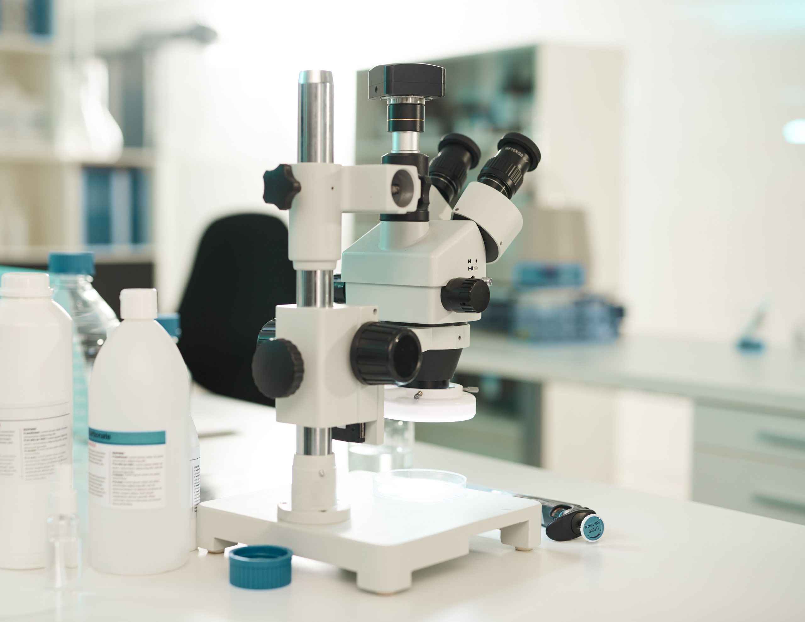
GoFertility — ranked as a top-50 American fertility clinic by Newsweek in 2023 — offers our patients cutting-edge technology using our in-office state-of-the-art equipment to produce optimal results. Since 2011 when GoFertility (or CRMRS) opened, we have bested the national pregnancy rate per embryo transfer every single year.
Dr. Saji Jacob — one of a select few dual board-certified infertility specialists in the country — and our professional team provide multiple high-tech treatments as part of our suite of fertility procedures to help couples have a baby. Those offerings include:
- In-vitro fertilization (IVF)
- Intracytoplasmic sperm injection (ICSI)
- Percutaneous epididymal sperm aspiration (PESA)
- Testicular sperm aspiration (TESA)
What is IVF and how does it work?
In-vitro fertilization paired with ICSI (intracytoplasmic sperm injection) is the industry standard for many infertility problems, including endometriosis, fibroids, ovulation disorders and male infertility. During IVF eggs are collected from the female patient’s ovaries at peak maturation. Meanwhile, a single, healthy sperm is collected from the male patient through the ICSI process (see more below).
Once retrieved, the mature eggs are injected with a single sperm and placed into incubators to allow for fertilization. They are then moved into the cryo bank for freezing for use in a future cycle when the embryo gets placed into the female’s uterus.
One full cycle of IVF-ICSI typically takes two to three weeks. The female patient will receive hormones for her specific situation to increase the number of eggs collected and timing of egg release. Hormone levels will then be checked via a series of blood samples. Ultrasound scans assist our team in knowing the right time for egg retrieval, with an injection causing the eggs to mature just prior to retrieval.
What are the risks of IVF-ICSI?
As with any assisted reproductive procedure, risks are present when undergoing IVF-ICSI though serious complications are very rare. If more than one embryo is placed in the female’s uterus, more than one baby — called a multiple pregnancy — can occur. The multiple pregnancy rate is higher with IVF-ICSI than a natural pregnancy, as twins account for about 20 percent of IVF-assisted pregnancies. Other IVF-related risks include:
- Ovarian hyper stimulation syndrome
- Reactions to medicine and anesthesia
- IVF cycle cancellation
The health of our patients is GoFertility’s chief concern. We seek to decrease the likelihood of a multiple pregnancy while maximizing the probability of a healthy pregnancy. While several factors — such as patient age and the number of times you have been through IVF-ICSI cycles — can impact this, we’ll guide you through the risks and help you make the best decision for your circumstances.
Every patient is different, and we won’t treat you like a number at GoFertility. Give our friendly and knowledgeable team a call to learn more about the in-vitro fertilization process.
What is ICSI and how does it work?
ICSI (intracytoplasmic sperm injection) promotes the highest chance of a successful pregnancy, with CRMRS’s team performing ICSI on all mature eggs selected for fertilization. With ICSI, our clinical laboratory embryologists select a single sperm and inject it into a single egg using a microscopic needle.
The benefit with this process is our team can catch the best sperm — healthy, oval heads with long, strong tails that aren’t broken — to increase the chances of successful fertilization. ICSI is particularly helpful for severe male infertility factors such as low sperm count, low sperm motility and high sperm abnormality.
ICSI has helped hundreds of GoFertility’s patients — not to mention couples around the world — successfully have a baby when other procedures were ineffective. Top-quality equipment, skilled personnel and attention to detail are required for a successful ICSI procedure, and you’ll receive all of that when you visit GoFertility.
What are PESA and TESA and how do they work?
PESA (percutaneous epididymal sperm aspiration) and TESA (testicular sperm aspiration) are complicated terms that simply mean gathering sperm from the testes when that process doesn’t happen naturally. These minimally invasive in-office procedures allow Dr. Jacob to collect sperm for the fertilization of eggs.
PESA and TESA are virtually painless, completed quickly and result in immediate recovery. The procedure takes about 20 to 40 minutes to complete in the privacy of our office. Once the sperm is gathered our CAP-certified lab uses an HOS test to identify live sperm, a process previously thought unimaginable.
GoFertility in Creve Coeur is always taking new patients, and we can typically set up a new patient appointment within a week. In addition to IVF-ICSI, PESA and TESA we offer the assisted hatching of embryos and donor eggs when necessary. We’ll find the best and safest solution for your needs to fulfill your dream of having a baby. Call us at 314-473-1285 or email us today to learn more!